Optical mapping of the pig heart in situ under artificial blood circulation
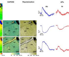
The emergence of optical imaging has revolutionized the investigation of cardiac electrical activity and associated disorders in various cardiac pathologies. The electrical signals of the heart and the propagation pathways are crucial for elucidating the mechanisms of various cardiac pathological conditions, including arrhythmia. The synthesis of near-infrared voltage-sensitive dyes and the voltage sensitivity of the FDA-approved dye Cardiogreen have increased the importance of optical mapping (OM) as a prospective tool in clinical practice. We aimed to develop a method for the high-spatiotemporal-resolution OM of the large animal hearts in situ using di-4-ANBDQBS and Cardiogreen under patho/physiological conditions. OM was adapted to monitor cardiac electrical behaviour in an open-chest pig heart model with physiological or artificial blood circulation. We detail the methods and display the OM data obtained using di-4-ANBDQBS and Cardiogreen. Activation time, action potential duration, repolarization time and conduction velocity maps were constructed. The technique was applied to track cardiac electrical activity during regional ischaemia and arrhythmia. Our study is the first to apply high-spatiotemporal-resolution OM in the pig heart in situ to record cardiac electrical activity qualitatively under artificial blood perfusion. The use of an FDA-approved voltage-sensitive dye and artificial blood perfusion in a swine model, which is generally accepted as a valuable pre-clinical model, demonstrates the promise of OM for clinical application.
Full article: https://www.ncbi.nlm.nih.gov/pmc/articles/PMC7244500/
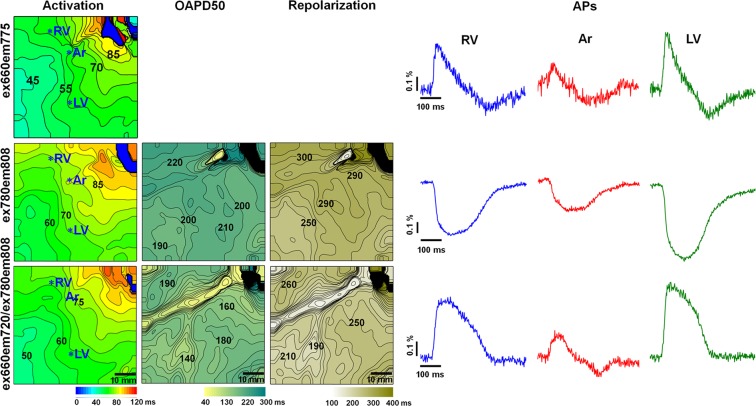